|
Case Report
Gaucher disease type 3 presenting with deformation of inferior limbs and bone infarction: A case report
1 Department of Radiology, Mother and Child Hospital, CHU Ibn-Sina, BP 6527, Rue Lamfadel Cherkaoui, Rabat, Morocco
Address correspondence to:
Zakaria Abide
Department of Radiology, Mother and Child Hospital, CHU Ibn-Sina, BP 6527, Rue Lamfadel Cherkaoui, Rabat,
Morocco
Message to Corresponding Author
Article ID: 100024R02ZA2023
Access full text article on other devices

Access PDF of article on other devices


How to cite this article
Abide Z, Es-Sebbani C, Cherraqi A, El Haddad S, Allali N, Chat L. Gaucher disease type 3 presenting with deformation of inferior limbs and bone infarction: A case report. Edorium J Radiol 2023;9(2):1–4.ABSTRACT
Introduction: Gaucher disease (GD) is a lysosomal storage disorder characterized by the accumulation of glucocerebroside in various cells throughout the body. Bone infarction is a common and fearsome complication.
Case Report: We present the case of a 5-year-old child diagnosed with Gaucher disease type 3, who exhibited deformation of the inferior limbs and bone pain. Upon evaluation, radiographic examination of the limbs revealed a characteristic triangular appearance of the metaphysis and a serpiginous sclerotic area. A magnetic resonance imaging was performed to confirm the diagnosis of bone infarction.
Conclusion: Gaucher disease is a complex genetic disorder. Bone involvement is a significant manifestation causing pain, bone crises, deformities. Various imaging techniques can help for an accurate diagnosis and for a timely intervention and prevention of the disease progression.
Keywords: Bone infraction, Gaucher disease, MRI, Standard radiography
INTRODUCTION
Gaucher disease is a rare genetic storage disorder characterized by a deficiency of the lysosomal enzyme glucocerebrosidase, leading to the accumulation of glucocerebroside in various cells throughout the body [1]. Bone complications in Gaucher disease can cause debilitating symptoms, including acute bone crises, chronic pain, spinal deformities, and joint destruction. Diagnosis often involves a thorough assessment of bone condition using imaging techniques such as X-rays, bone scintigraphy, and magnetic resonance imaging (MRI). Management of bone infarction, a common complication, focuses on treating the underlying metabolic disorder through enzyme replacement therapy and substrate reduction therapy [2].
CASE REPORT
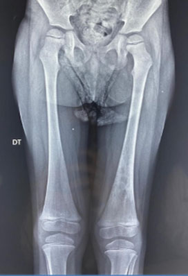
We report the case of a 5-year-old child with a known medical history of Gaucher disease type 3, who presented with deformities of the inferior limbs and complaints of bone pain. Upon evaluation, radiographic examination of the limbs revealed a characteristic triangular appearance of the metaphyses, with an indistinct boundary between the cortex and medulla, consistent with the Erlenmeyer flask deformity (Figure 1).
Additionally, during the assessment, an incidental finding of a serpiginous sclerotic area was noted in the lower left femoral metaphysis, raising suspicion of bone infraction.
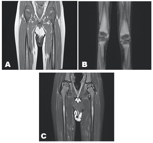
To gain further insight into the extent and nature of the skeletal abnormalities, magnetic resonance imaging (MRI) was performed. The MRI scans demonstrated multiple bilateral signal anomalies in the diaphyseal and metaphyseal regions of the femurs. These anomalies appeared as areas of low signal intensity on T1-weighted images and exhibited high signal intensity on short tau inversion recovery (STIR) images. Notably, the infarcted zones were delineated by a map-like serpiginous border, serving as a distinct visual marker of the affected areas (Figure 2A, Figure 2B, Figure 2C).
DISCUSSION
Gaucher disease is a storage disorder caused by a deficiency of the lysosomal enzyme glucocerebrosidase. It is characterized by the accumulation of glucocerebroside in the cells of the reticuloendothelial system (liver, spleen, bone marrow) and lymph nodes, primarily affecting macrophages and transforming them into Gaucher cells. Gaucher cells are found in the splenic sinuses, replacing Kupffer cells in the liver, alveolar macrophages in the lungs, and infiltrating the bone marrow. The mode of transmission is autosomal recessive [1].
The pathophysiology of bone involvement is only partially understood at present. The presence of overloaded macrophages within the bone marrow leads to an increase in intra-medullary pressure. This elevated pressure causes cortical thinning on one hand and stimulates osteoclastic resorption on the other hand, resulting in a decoupling of bone remodeling. This condition specifically affects types 1 and 3 and appears to be favored by splenectomy. Understanding this condition is crucial as it may be the initial manifestation of the disease, and its diagnosis will enable the implementation of specific treatment to prevent its progression. It is a debilitating complication: the pain can be acute and sudden, referred to as bone crises, often corresponding to bone infarction, and sometimes to a fracture. Alternatively, the pain can be more moderate and chronic, associated with spinal deformity or secondary joint destruction due to epiphyseal osteonecrosis [2].
During the diagnosis of MG (presumably referring to a medical condition), it is recommended to carefully assess the bone condition. This evaluation will help detect any previous infarction-related sequelae that could be treated in the case of secondary osteoarthritis. It also provides a baseline imaging assessment that will be useful in the event of future complications.
The following examinations are recommended at the time of diagnosis:
- Standard X-rays of the pelvis, spine, femurs, tibias, and humerus.
- Technetium 99m bone scintigraphy to identify any hyperfixation of bone lesions.
- Magnetic resonance imaging of the spine, pelvis, and lower limbs, including any hyperfixation areas identified in the bone scintigraphy. This will allow quantification of the extent of bone infiltration and determine whether the lesions are recent or old.
- Computed tomography scans are not recommended as the first-line imaging technique due to radiation exposure.
In general, it is used T1-weighted and T2-weighted sequence [1],[2].
Bone involvement is a recognized feature of Gaucher disease and can lead to significant morbidity. The Erlenmeyer flask deformity, observed in the radiographic examination, is a classic finding attributed to abnormal bone remodeling due to the infiltration of glucocerebroside-laden macrophages. This deformity often occurs in long bones and is characterized by the widening of the metaphyses with a characteristic triangular appearance [3].
Bone infarction, also known as avascular necrosis or osteonecrosis, results from impaired blood supply to the affected bone, leading to ischemic necrosis and subsequent structural damage. It is believed to occur due to the obstruction of blood vessels by the accumulation of glucocerebroside-laden macrophages [1].
The management of bone infarction in Gaucher disease primarily focuses on the treatment of the underlying metabolic disorder. Enzyme replacement therapy and substrate reduction therapy are the mainstays of treatment for Gaucher disease, aiming to reduce the accumulation of glucocerebroside and alleviate systemic manifestations. While these treatments may not directly target bone infarction, they have been shown to improve overall disease control and potentially slow the progression of skeletal complications [3].
CONCLUSION
Gaucher disease is a complex genetic disorder characterized by the deficiency of the lysosomal enzyme glucocerebrosidase. Bone involvement is a significant manifestation causing pain, bone crises, deformities. Various imaging techniques can help for an accurate diagnosis and for a timely intervention and prevention of the disease progression.
REFERENCES
1.
Lavigne C, Bergelin-Besancon A, Maillot F. Les manifestations osseuses de la maladie de Gaucher. Réalités en rhumatologie. 2010. [Available at: https://www.realites-cardiologiques.com/2010/05/31/les-manifestations-osseuses-de-la-maladie-de-gaucher/]

2.
Hughes D, Mikosch P, Belmatoug N, et al. Gaucher disease in bone: From Pathophysiology to practice. J Bone Miner Res 2019;34(6):996–1013. [CrossRef]
[Pubmed]
3.
Bembi B, Ciana G, Mengel E, Terk MR, Martini C, Wenstrup RJ. Bone complications in children with Gaucher disease. Br J Radiol 2002;75 Suppl 1:A37–44. [CrossRef]
[Pubmed]
SUPPORTING INFORMATION
Author Contributions
Zakaria Abide - Conception of the work, Design of the work, Acquisition of data, Analysis of data, Drafting the work, Revising the work critically for important intellectual content, Final approval of the version to be published, Agree to be accountable for all aspects of the work in ensuring that questions related to the accuracy or integrity of any part of the work are appropriately investigated and resolved.
Chaimae Es-Sebbani - Conception of the work, Design of the work, Analysis of data, Revising the work critically for important intellectual content, Final approval of the version to be published, Agree to be accountable for all aspects of the work in ensuring that questions related to the accuracy or integrity of any part of the work are appropriately investigated and resolved.
Amine Cherraqi - Analysis of data, Revising the work critically for important intellectual content, Final approval of the version to be published, Agree to be accountable for all aspects of the work in ensuring that questions related to the accuracy or integrity of any part of the work are appropriately investigated and resolved.
Siham El Haddad - Revising the work critically for important intellectual content, Final approval of the version to be published, Agree to be accountable for all aspects of the work in ensuring that questions related to the accuracy or integrity of any part of the work are appropriately investigated and resolved.
Nazik Allali - Revising the work critically for important intellectual content, Final approval of the version to be published, Agree to be accountable for all aspects of the work in ensuring that questions related to the accuracy or integrity of any part of the work are appropriately investigated and resolved.
Latifa Chat - Revising the work critically for important intellectual content, Final approval of the version to be published, Agree to be accountable for all aspects of the work in ensuring that questions related to the accuracy or integrity of any part of the work are appropriately investigated and resolved.
Guaranter of SubmissionThe corresponding author is the guarantor of submission.
Source of SupportNone
Consent StatementWritten informed consent was obtained from the patient for publication of this article.
Data AvailabilityAll relevant data are within the paper and its Supporting Information files.
Conflict of InterestAuthors declare no conflict of interest.
Copyright© 2023 Zakaria Abide et al. This article is distributed under the terms of Creative Commons Attribution License which permits unrestricted use, distribution and reproduction in any medium provided the original author(s) and original publisher are properly credited. Please see the copyright policy on the journal website for more information.