|
Case Report
Chilaiditi sign secondary to a right renal agenesis
1 Resident in Radiology, Department of Radiology, National Institute of Oncology, UHC Ibn Sina, Rabat, Morocco
2 Professor Chief of the Radiology, Department Oncology, National Institute UHC Ibn Sina, Rabat, Morocco
3 Assistan professor, Department of Radiology, National Institute of Oncology, UHC Ibn Sina, Rabat, Morocco
Address correspondence to:
Kenza Berrada
Rabat 140000,
Morocco
Message to Corresponding Author
Article ID: 100023R02KB2022
Access full text article on other devices

Access PDF of article on other devices


How to cite this article
Berrada K, El Bakkari A, Jerguigue H, Latib R, Omor Y. Chilaiditi sign secondary to a right renal agenesis. Edorium J Radiol 2022;8(2):10–13.ABSTRACT
Chilaiditi’s sign is a rare radiological finding in which a portion of the colon or small intestine is interposed between the liver and right hemidiaphragm. Associated to pain, it is called Chilaiditi syndrome. We describe the case of a patient who was admitted to our radiology department to release a body-computed tomography (CT) for its breast cancer extension assessment. We incidentally discovered a colic hepato-phrenic interposition related to a Chilaiditi sign associated with right renal agenesis. In this case report we treated Chilaiditi sign can exist without Chilaiditi syndrome, as it could be associated with other pathologies and abnormal anatomic malformation like renal agenesis.
Keywords: Chilaiditi sign, Chilaiditi syndrome, Renal agenesis
INTRODUCTION
Chilaiditi sign is defined by hepatophrenic intestinal interposition. When it is associated to pain, it is called Chilaiditi syndrome; commonly, incidentally discovered, and/or secondary to a complication like intestinal occlusion and bowel ischemia. We describe the case of a patient who was admitted to our radiology department to release a body-computed tomography (CT) for its breast cancer extension assessment, we incidentally discovered a colic hepato-phrenic interposition related to a Chilaiditi sign associated with right renal agenesis.
CASE REPORT
A 66-year-old female with a newly diagnosed breast carcinoma presented with right upper quadrant pain and sensation of heaviness.
Investigations
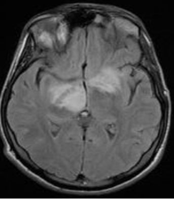
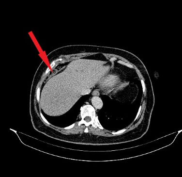
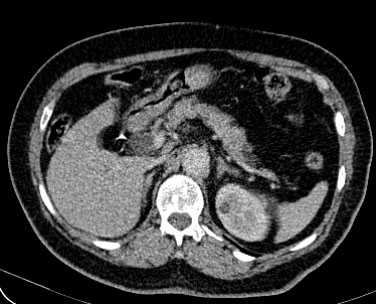
A contrast-enhanced CT of thoracic, abdomen, and pelvis was performed with series obtained in portal phase for initial extension assessment and to explore the symptomatology. It demonstrates an interposition of transverse colon between liver and diaphragm (Figure 1). Besides CT also shows a right empty renal fossa with left normal kidney and no other anomalies were noted, related to a renal agenesis (Figure 2).
Differential diagnosis
In front of the medical history of breast carcinoma the diagnosis of an interposed intestine between the diaphragm and the liver can be mistaken with a peritoneal node, especially when it is empty. Pneumoperitoneum can be another misleading diagnosis in a traumatic context. Finally, a perihepatic abscess can mimic it.
Treatment
No intervention is required for an asymptomatic patient with Chilaiditi sign. Those with abdominal pain or distension are usually treated conservatively with analgesia and fluid resuscitation. Patients with recurrent presentations or evidence of bowel ischemia may be offered surgical treatment.
DISCUSSION
Chilaiditi syndrome is used to describe pain caused by an interposition of the intestine between liver and diaphragm, which is called Chilaiditi sign. Firstly, described in 1910, it is named after a Greek radiologist Chilaiditi Demetrius [1].
It is an uncommon syndrome, occurring its incidence varies from 0.25% to 0.28% of the general population and affects mainly male and elderly patients [2], [3].
Different predisposing factors engender the development of Chilaiditi sign [4]. Its cause still remains unknown, but it is probably multifactorial. Congenital anatomical abnormalities such as colonic elongation and laxity of colonic and hepatic suspensory ligaments have been postulated as the principal predisposing factors. Other predisposing situations can be acquired such as an enlarged lower thoracic outlet (e.g., pregnancy, emphysema, and cirrhosis with ascites), or organ shrinkage (atrophic cirrhosis of the liver). Fat deposition between the liver and the colon, in obese individuals, widens the space between the two organs and can also favor the migration of the colon [5].
It is usually asymptomatic and mostly discovered incidentally on CT examination. Most frequent symptoms are abdominal pain, nausea, vomiting, and respiratory distress. Differential diagnoses of this radiographic sign include pneumoperitoneum, peritoneal nodes, and subphrenic abscess. Main complications are intestinal occlusion and bowel ischemia [6]. Chilaiditi syndrome has been associated with a variety of pulmonary or gastrointestinal malignancies (involving the colon, rectum, or stomach) [7].
If Chilaiditi sign is visualized and the patient is symptomatic, it is then referred to as Chilaiditi’s syndrome. Gastrointestinal symptoms, such as epigastric pain, nausea, vomiting, constipation, abdominal distention are the main symptoms [6].
Diagnosis of the disease is made primarily through CT scans. It demonstrates the air below the diaphragm associated with visible intestinal haustra, which does not change with variation in patient position. Besides, elevated right hemidiaphragm above the liver by the intestine and depression of the superior margin of the liver below the left hemidiaphragm are associated. The differential diagnoses of Chilaiditi syndrome can include diaphragmatic hernia, pneumoperitoneum, and subphrenic abscess [7].
Complications of Chilaiditi syndrome may include a volvulus of the cecum, splenic flexure, or transverse colon. Cecal perforation and, rarely, perforated subdiaphragmatic appendicitis can also occur as complications of Chilaiditi syndrome [8]. Respiratory distress, cardiac arrhythmia and serious complications, can also be seen [9].
CONCLUSION
In this case report we treated Chilaiditi sign can exist without Chilaiditi syndrome, as it could be associated with other pathologies and abnormal anatomic malformation like renal agenesis. Radiologic investigation is crucial to stand up the right diagnosis, the radiologist has to pay attention to not confuse it with other differential diagnosis, mainly with pneumoperitoneum and peritoneal nodes for patients with malignancies.
REFERENCES
1.
Maizlin ZV. Wonders of Radiology. USA: CreateSpace Independent Publishing Platform; 2010.

2.
McNamara RF, Cusack S, Hallihan P. Chilaiditi’s Syndrome. West J Emerg Med 2009;10(4):250.
[Pubmed]
3.
Chen SY, Liu CT, Tsai YC, Yu JC, Lin CH. Sigmoid volvulus associated Chiladiti’s syndrome. Rev Esp Enferm Dig 2007;99(8):482–3. [CrossRef]
[Pubmed]
4.
Tangri N, Singhal S, Sharma P, et al. Coexistence of pneumothorax and Chilaiditi sign: A case report. Asian Pac J Trop Biomed 2014;4(1):75–7. [CrossRef]
[Pubmed]
5.
Sorrentino D, Bazzocchi M, Badano L, Toso F, Giagu P. Heart-touching Chilaiditi’s syndrome. World J Gastroenterol 2005;11(29):4607–9. [CrossRef]
[Pubmed]
6.
Antonacci N, Di Saverio S, Biscardi A, Giorgini E, Villani S, Tugnoli G. Dyspnea and large bowel obstruction: A misleading Chilaiditi syndrome. Am J Surg 2011;202(5):e45–7. [CrossRef]
[Pubmed]
7.
Kapania EM, Link C, Eberhardt JM. Chilaiditi syndrome: A case report highlighting the intermittent nature of the disease. Case Rep Med 2018;2018:3515370. [CrossRef]
[Pubmed]
8.
Moaven O, Hodin RA. Chilaiditi syndrome: A rare entity with important differential diagnoses. Gastroenterol Hepatol (N Y) 2012;8(4):276–8.
[Pubmed]
9.
Özer C, Zenger S. Chilaiditi syndrome in a patient with urological problems: Incidental diagnosis on computed tomography. Can Urol Assoc J 2012;6(2):E75–6. [CrossRef]
[Pubmed]
SUPPORTING INFORMATION
Author Contributions
Kenza Berrada - Conception of the work, Design of the work, Acquisition of data, Analysis of data, Drafting the work, Final approval of the version to be published, Agree to be accountable for all aspects of the work in ensuring that questions related to the accuracy or integrity of any part of the work are appropriately investigated and resolved.
Asaad El Bakkari - Drafting the work, Revising the work critically for important intellectual content, Final approval of the version to be published, Agree to be accountable for all aspects of the work in ensuring that questions related to the accuracy or integrity of any part of the work are appropriately investigated and resolved.
Hounayda Jerguigue - Revising the work critically for important intellectual content, Final approval of the version to be published, Agree to be accountable for all aspects of the work in ensuring that questions related to the accuracy or integrity of any part of the work are appropriately investigated and resolved.
Rachida Latib - Revising the work critically for important intellectual content, Final approval of the version to be published, Agree to be accountable for all aspects of the work in ensuring that questions related to the accuracy or integrity of any part of the work are appropriately investigated and resolved.
Youssef Omor - Revising the work critically for important intellectual content, Final approval of the version to be published, Agree to be accountable for all aspects of the work in ensuring that questions related to the accuracy or integrity of any part of the work are appropriately investigated and resolved.
Guaranter of SubmissionThe corresponding author is the guarantor of submission.
Source of SupportNone
Consent StatementWritten informed consent was obtained from the patient for publication of this article.
Data AvailabilityAll relevant data are within the paper and its Supporting Information files.
Conflict of InterestAuthors declare no conflict of interest.
Copyright© 2022 Kenza Berrada et al. This article is distributed under the terms of Creative Commons Attribution License which permits unrestricted use, distribution and reproduction in any medium provided the original author(s) and original publisher are properly credited. Please see the copyright policy on the journal website for more information.