|
Case Report
Meningo-encephalo-vasculitis, optic neuritis, and thrombotic complications: About a fulminant mucormycosis in a diabetic patient
1 Resident in Radiology, Neuroradiology Department, Specialty Hospital of Rabat, Morocco
2 Resident, Mycology Department, IBN SINA Hospital Center, Rabat, Morocco
3 Professor in Radiology, Neuroradiology Department, Specialty Hospital of Rabat, Morocco
Address correspondence to:
Ibtissam El Ouali
Resident in Radiology, Neuroradiology Department, Specialty Hospital of Rabat, Avenue Lalla Asmaa, AL AZHAR Residency, Salé,
Morocco
Message to Corresponding Author
Article ID: 100020R02IO2022
Access full text article on other devices

Access PDF of article on other devices

How to cite this article
El Ouali I, Hamzaoui A, Diallo ID, Fikri M, Jiddane M, Touarsa F. Meningo-encephalo-vasculitis, optic neuritis, and thrombotic complications: About a fulminant mucormycosis in a diabetic patient. Edorium J Radiol 2022;8(1):1–5.ABSTRACT
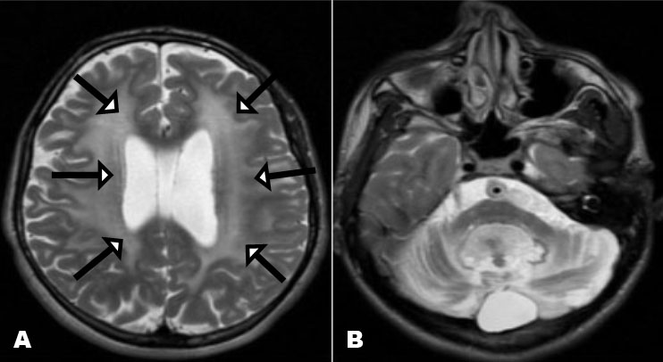
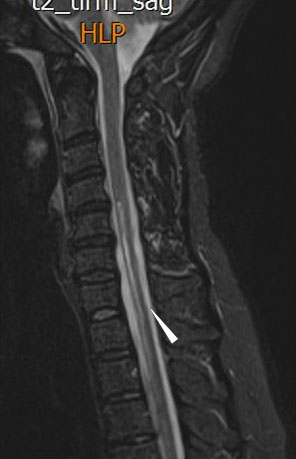
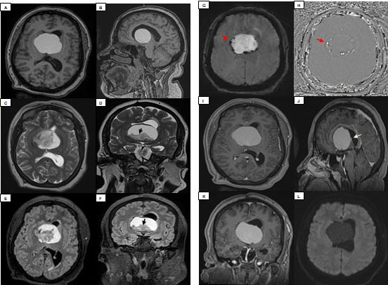
Mucormycosis is a destructive, potentially fatal, and opportunistic fungal infection caused by filamentous Mucorales which commonly affect immunocompromised hosts. This infection might take different forms such as gastrointestinal, pulmonary, cutaneous or even a disseminated form, yet the rhinocerebral localization is historically the primary presentation of the disease and most common type. It originates in the nasal mucosa owing to fungal inoculation, then it spreads through paranasal sinuses and orbits to the brain and its vessels especially the cavernous sinus, leading to thrombotic complications including arterial thrombosis. Herein, we present a case of a 35-year-old male with poorly controlled diabetes who presented with decompensated diabetes, in whom the clinical examination finds subtle signs of orbital cellulitis. The patient subsequently had worsening necrotizing orbital cellulitis which required surgical drainage of the left ethmoid along with large spectrum antibiotic therapy; this was complicated by the development of meningo-encephalo-vasculitis as well as cavernous sinus and left internal carotid thrombosis. Tissue cultures revealed evidence of Rhizopus.
Keywords: CT scan, Diabetes, MRI, Mucormycosis
SUPPORTING INFORMATION
Acknowledgments
I would like to express my gratitude to my professors and all the colleagues who participated in the completion of this work. The authors declare that they have no competing interests.
Author ContributionsIbtissam El Ouali - Substantial contributions to conception and design, Acquisition of data, Analysis of data, Interpretation of data, Drafting the article, Final approval of the version to be published
Abdeljalil Hamzaoui - Acquisition of data, Drafting the article, Final approval of the version to be published
Ibrahima Dokal Diallo - Acquisition of data, Drafting the article, Final approval of the version to be published
Meriem Fikri - Substantial contributions to conception and design, Drafting the article, Final approval of the version to be published
Mohamed Jiddane - Substantial contributions to conception and design, Drafting the article, Final approval of the version to be published
Firdaous Touarsa - Substantial contributions to conception and design, Revising it critically for important intellectual content, Final approval of the version to be published
Guaranter of SubmissionThe corresponding author is the guarantor of submission.
Source of SupportNone
Consent StatementWritten informed consent was obtained from the patient for publication of this article.
Data AvailabilityAll relevant data are within the paper and its Supporting Information files.
Conflict of InterestAuthors declare no conflict of interest.
Copyright© 2022 Ibtissam El Ouali et al. This article is distributed under the terms of Creative Commons Attribution License which permits unrestricted use, distribution and reproduction in any medium provided the original author(s) and original publisher are properly credited. Please see the copyright policy on the journal website for more information.