 |
|
Case Report
| ||||||
| Huge renal angiomyolipoma with multiple aneurysms treated with staged transcatheter arterial embolization using N-butyl cyanoacrylate and microcoils: A case report | ||||||
| Zenjiro Sekikawa1, Toh Yamamoto1, Ryo Aoki1, Shintaro Furugori2, Shigeo Takebayashi1 | ||||||
|
1Department of Radiology, Yokohama City University Medical Center, Yokohama, Kanagawa, Japan 2Department of Critical and Emergency Center, Yokohama City University Medical Center, Yokohama, Kanagawa, Japan | ||||||
| ||||||
|
[HTML Abstract]
[PDF Full Text]
[Print This Article] [Similar articles in PubMed] [Similar articles in Google Scholar] |


| How to cite this article |
| Sekikawa Z, Yamamoto T, Aoki R, Furugori S, Takebayashi S. Huge renal angiomyolipoma with multiple aneurysms treated with staged transcatheter arterial embolization using N-butyl cyanoacrylate and microcoils: A case report. Edorium J Radiol 2018;4:100010R02ZS2018. |
|
ABSTRACT
| ||||||
|
Introduction: We report a rare case of a huge bleeding renal angiomyolipoma (AML) with multiple aneurysms treated with staged transcatheter arterial embolization (TAE) using N-butyl cyanoacrylate (NBCA) and additional microcoils. After three TAEs, we succeeded in managing hemorrhage and aneurysms. We also gained 50% tumor regression with preserved viable renal parenchyma. Case Report: A 37-year-old female was admitted at our emergency center due to retroperitoneal hemorrhage. Computed tomography (CT) revealed a huge right renal AML of 26 cm in diameter with multiple aneurysms bleeding in the retroperitoneal space. An emergent TAE was performed, and we managed bleeding by using NBCA instantly, but large aneurysms in the tumor vessel remained. We planned staged TAE for the aneurysms left at the proximal side of the right renal artery. We used detachable coils as embolizing agents after NBCA, and the second TAE was completed uneventfully. However, follow-up CT examination revealed another hemorrhage, the third TAE was performed. An arteriogram of the right renal artery revealed tumor stain from other branches of the renal artery, which we did not embolize before. These branches were embolized completely using NBCA. Follow-up CT examination showed the aneurysms stopped and a tumor regression of about 50% in diameter. Conclusion: TAE using NBCA is very useful for treating renal AML because it can provide promising effects and low complications with a minimally invasive technique. We choose NBCA as a first-choice embolizing agent for treating renal AML even if it is huge and has aneurysm with staged TAE. Keywords: Aneurysm, Embolization, Hemorrhage, NBCA, Renal angiomyolipoma | ||||||
|
INTRODUCTION
| ||||||
|
Transcatheter arterial embolization (TAE) has become the primary treatment in the management of renal angiomyolipoma (AML) [1], [2]. We report a rare case of a huge bleeding renal AML with multiple aneurysms treated with repeated TAE using N-butyl cyanoacrylate (NBCA) and additional microcoils. After three TAEs, we succeeded in managing hemorrhage and aneurysms and gaining 50% tumor regression with consistent viable renal parenchyma. | ||||||
|
CASE REPORT
| ||||||
|
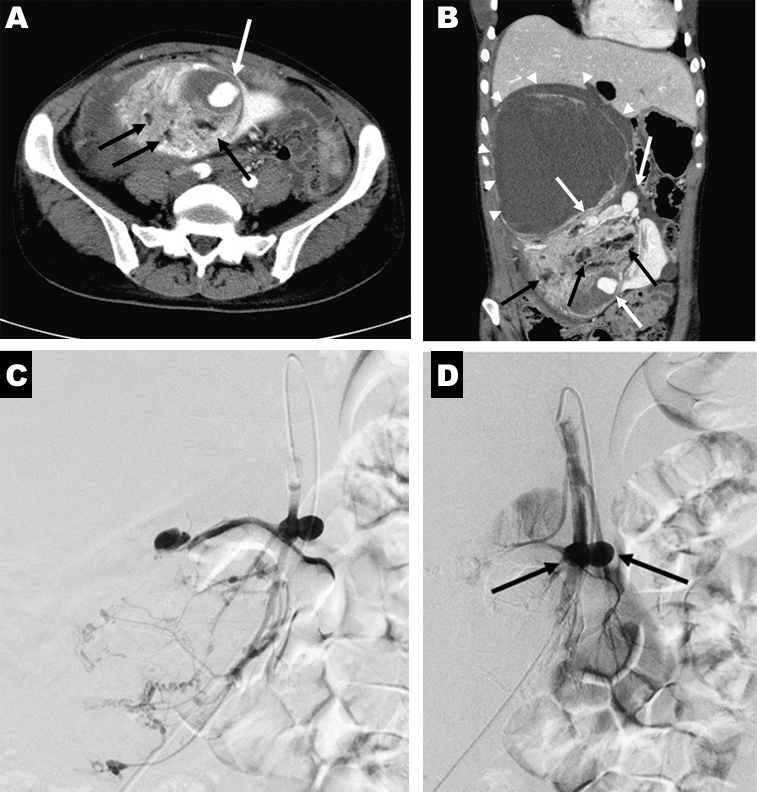
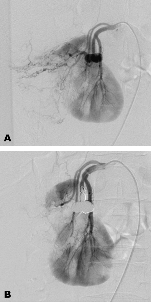
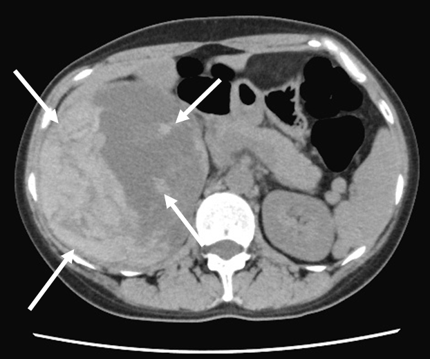
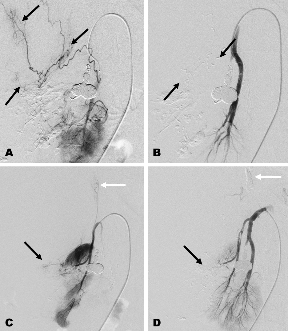
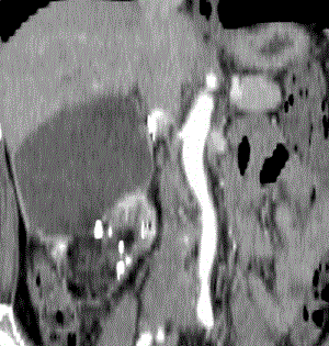
A 37-year-old female was referred to the Department of critical and emergency center in our hospital because of acute right flank pain. Computed tomography (CT) revealed a huge right renal tumor of 26 cm in diameter with multiple aneurysms bleeding in the retroperitoneal space (Figure 1 A and B). The tumor contained a small amount of fat and soft tissue density, which was thought to be angiomyomatous portion in the caudal part of the tumor, and large cystic area in the cranial part of the tumor, which was diagnosed as renal angiomyolipoma (AML). After the patient was given an initial treatment, an emergent transcatheter artery embolization (TAE) was planned to stop the bleeding. We confirmed that the legal representatives of the patient were given a comprehensive written statement of information about the treatment, including information on TAE, and their consent was documented in the clinical record. The patient’s history was unremarkable, and she had no history of tuberous sclerosis complex (TSC). A right renal artery angiogram was obtained using a 5 French (F) shepherd hook catheter (Catex-CX; Gadelius Medical, Tokyo, Japan). The angiogram showed large tumor stain with multiple aneurysms, though whole estimation was insufficient due to its high flow. A 5.2 F cobra-head occlusion catheter (Selecon MP catheter; Terumo, Tokyo, Japan) was chosen to decrease the main flow and enable us to see the details of the tumor’s vessels. Under inflation of the balloon of the catheter, the detail of the vessels inside the tumor was revealed clearly. There were three main branches from the proximal portion of the right renal artery trunk, and multiple aneurysms all seemed to originate from the central branch of these three branches (Figure 1 C). A 2.0 F microcatheter (Bobsled; Kaneka Medical, Osaka, Japan) was inserted into the distal portion of the central branch, a total of 2 ml of N-butyl cyanoacrylate (NBCA) (Histoacryl B; Braun, Melsungen, Germany) mixed with iodized oil (Lipiodol; Terumo) at a ratio of 1:4 (20%) was infused through it. Two large aneurysms remained in the proximal portion of this branch (Figure 1 D). Treatment for these aneurysms was decided to be performed the next time because the initial treatment for the bleeding was thought to be enough at the time, and it had taken little time to perform TAE. Further, it seemed to need a more deliberate coil embolization technique for the aneurysms left at the proximal side of the branch. The next TAE was planned after two months when the patient was completely recovered from her initial unstable situation. When the next TAE was just about to begin, she reported chest discomfort, which gradually became vital shock, which was later revealed to be a latex allergy. Therefore, the next TAE was scheduled under general anesthesia. A follow-up CT examination showed the tumor had regressed to 18 cm in diameter, but two aneurysms were still left inside the tumor. The second TAE was performed five months after the first TAE with the patient under general anesthesia. Using a 4 F cobra-head catheter (Terumo), a 2 F microcatheter (Bobsled; Kaneka Medical) was inserted into the central branch that had aneurysms, and an arteriogram was obtained (Figure 2 A). The arteriogram showed tumor stain remained in the distal part of the branch; hence, a small amount of NBCA mixed with iodized oil at a ratio of 25% (1:3) was infused through the microcatheter. Subsequently, a 2.6 F microcatheter (PX Slim; Penumbra; Alameda, CA, USA) was inserted into the proximal portion of the same branch, where two aneurysms were cited. Seven detachable microcoils (Ruby coil; Penumbra) ranging from 16 mm to 6 mm in diameter were placed inside the aneurysms, and total packing of the aneurysm was completed. A right renal arteriogram confirmed occlusion of these aneurysms, and no residual aneurysm and tumor stain was observed (Figure 2 B). Four days after second TAE, the anemia was still present; hence, a CT examination was performed, which showed heterogeneous high-density area in the cranial portion of the cystic area of the tumor, suggesting new active bleeding (Figure 3). There was no apparent extravasation after administration of the contrast media, but additional TAE was considered. The third TAE was performed two days after the CT examination with the patient under general anesthesia. A 4 F cobra-head catheter was inserted into the right renal artery, and an arteriogram was obtained. Two branches, which were still not embolized, showed a small amount of tumor stain. A 1.7 F microcatheter (progreat λ; Terumo) was inserted into the distal branch from the lower branch, and a small amount of NBCA mixed with iodized oil at a ratio of 10% (1:9) was infused through the microcatheter (Figure 4 A, B). The arteriogram of the upper branch showed two branches from the distal portion of this artery; one towards the cranial and the other towards the lateral side of the tumor, both of which had tumor stain (Figure 4 C). These arteries were embolized with 10% NBCA. Finally, complete occlusion of these arteries was confirmed through the right renal arteriogram (Figure 4 D). No apparent complication was observed during and after the procedure. After the third TAE, the patient was discharged from the hospital uneventfully, and the follow-up reconstructed CT examination performed four months later showed extinguishment of the aneurysms and tumor regression of about 50% in diameter with preservation of the normal renal parenchyma (Figure 5). The patient’s renal function is within normal limits. The preoperative serum creatinine level was 0.42 mg/dl, and at last follow-up was 0.54 mg/dl. | ||||||
|
| ||||||
|
| ||||||
| ||||||
|
| ||||||
| ||||||
DISCUSSION | ||||||
|
Renal AML is the most common benign renal tumor, accounting for 0.3% to 3% of all renal masses [3]. They include fat, blood vessels, and smooth muscle in various proportions. They are divided into sporadic and non-sporadic groups [4]. The sporadic group accounts for approximately 80% of the incidences, which occur mainly in middle-aged females, and the lesion is usually solitary. The non-sporadic group is associated with TSC, an autosomal-dominant multiorgan phacomatosis predisposing to benign tumor such as renal AML [5]. In the setting of TSC, renal AML is often larger, multiple, bilateral, and symptomatic [6]. One of the most dreadful events associated with this tumor is bleeding, which may become life-threatening. The bleeding tendency is thought to be from the tumor’s angiogenic component, of which the vessel walls are abnormal with no internal elastic lamina. Further, the smooth muscle of the vessel is replaced by fibrous tissue, making the vessels rigid, tortuous, and prone to aneurysms [7]. Reports have suggested that hemorrhagic presentation was significantly more frequent in patients with intralesional aneurysms [8], [9]. TAE is currently becoming the first treatment of choice for renal AML because it is less invasive and has less adverse effects and can conserve the renal function [1]. Additionally, TAE can be used not only for active bleeding, but also as a prophylactic method and preoperative adjunct treatment to reduce intraoperative hemorrhage [1],[6]. Some authors reported that TAE for AML has less postoperative morbidity than partial nephrectomy, with shorter hospitalization [10], [11] . Various embolized agents were reportedly used for TAE of the renal AML, such as gelatin sponge, microbeads, ethanol, polyvinyl alcohol particles, coils, NBCA, and a combination of these materials, though NBCA seems to not be popular among them. We used mainly NBCA as embolic material because it works instantly and correctly, providing an eternal promising effect on the target vessels. Additionally, it is easy to control its concentration and visualize the flow when it is mixed with iodized oil (lipiodol) [12]. Bardin et al. also emphasized the benefit of the usage of NBCA for TAE of the renal AML [8]. TAE was repeated three times consequently in this case. Bishay et al. reported that if the AML was large or had multiple feeding vessels, embolization was staged to keep complication rates low [2]. We encountered post embolization syndrome as a side effect, which manifests as fever, pain, nausea, and vomiting, but no severe complication occurred. Normal renal parenchyma and renal function were preserved after TAE. The tumor regression rate in the present case was about 50%. The rate was much higher than that reported in the previous published data, at around 25% [13]; the reasons might contribute to the use of NBCA and the rich amount of angiomyomatous component of the tumor in this case might cause this good result. | ||||||
|
CONCLUSION
| ||||||
|
TAE using NBCA is very useful for treating renal AML because it can provide a promising effect and low complications with minimal invasive technique. We choose NBCA as the first choice of the embolizing agent for treating renal AML even if it is huge and has aneurysm with staged TAE. | ||||||
|
REFERENCES
| ||||||
| ||||||

|
[HTML Abstract]
[PDF Full Text]
|
|
Author Contributions
Zenjiro Sekikawa – Substantial contributions to conception and design, Acquisition of data, Analysis and interpretation of data, Drafting the article, Revising it critically for important intellectual content, Final approval of the version to be published Toh Yamamoto – Substantial contributions to conception and design, Acquisition of data, Analysis and interpretation of data, Drafting the article, Final approval of the version to be published Ryo Aoki – Substantial contributions to conception and design, Acquisition of data, Analysis and interpretation of data, Drafting the article, Final approval of the version to be published Shintaro Furugori – Substantial contributions to conception and design, Acquisition of data, Analysis and interpretation of data, Drafting the article, Final approval of the version to be published Shigeo Takebayashi – Substantial contributions to conception and design, Analysis and interpretation of data, Drafting the article, Revising it critically for important intellectual content, Final approval of the version to be published |
|
Guarantor of Submission
The corresponding author is the guarantor of submission. |
|
Source of Support
None |
|
Consent Statement
Written informed consent was obtained from the patient for publication of this case report. |
|
Conflict of Interest
Author declares no conflict of interest. |
|
Copyright
© 2018 Zenjiro Sekikawa et al. This article is distributed under the terms of Creative Commons Attribution License which permits unrestricted use, distribution and reproduction in any medium provided the original author(s) and original publisher are properly credited. Please see the copyright policy on the journal website for more information. |
|
|