 |
|
Case Report
| ||||||
| A rare case report of acquired pericecal hernia complicating gastrointestinal endoscopy: Multidetector computed tomography findings revisited | ||||||
| Mohamed Ragab Nouh1, Ahmed Bassim Doma2, Mohamed Ahmed Yousef Elshazly3, Elsanousi Ibrahim Sabir4 | ||||||
|
1Department of Radiology, Faculty of Medicine, Alexandria University, Armed Force Hospital, King Abdualaziz Airbase, Dhahran, Kingdom of Saudi Arabia
2Department of Radiology, Armed Force Hospital, King Abdualziz Airbase, Dhahran, Kingdom of Saudi Arabia 3Department of Radiology, Faculty of Medicine, Al-Azhar University, Armed Force Hospital, King Abdualziz Airbase, Dhahran, Kingdom of Saudi Arabia 4Department of Surgery, Armed Force Hospital, King Abdualziz Airbase, Dhahran, Kingdom of Saudi Arabia | ||||||
| ||||||
|
[HTML Abstract]
[PDF Full Text]
[Print This Article] [Similar article in Pumed] [Similar article in Google Scholar] |


| How to cite this article |
| Nouh MR, Doma AB, Elshazly MAY, Sabir EI. A rare case report of acquired pericecal hernia complicating gastrointestinal endoscopy: Multidetector computed tomography findings revisited. Edorium J Radiol 2018;4:1–5. |
|
ABSTRACT
|
|
Introduction:
Internal hernia is an infrequent cause of intestinal obstruction. It represents a diagnostic challenge to both clinicians and radiologists. Recognizing internal hernia as an underlying cause of intestinal obstruction is both challenging and critical to avoid associated morbidities. Currently, multidetector computed tomography (MDCT) is pivotal in the diagnostic workup of acute abdomen, especially those cases presented by intestinal obstruction.
Case Report: A 69-year-old male was presented with acute abdominal pain and distension following combined upper and lower gastrointestinal screening endoscopy done to investigate recurrent nausea, dyspepsia and episodic vomiting. His medical history was relevant only for a previous appendectomy thirty years ago. Physical examination revealed abdominal distension, right iliac fossa tenderness and hyperactive bowel sounds and a provisional diagnosis of intestinal obstruction was instituted. Plain radiography showed no significant air-fluid levels while contrast-enhanced computed tomography of the abdomen revealed locally crowded fluid-filled dilated ileal segments within the right lower abdominal quadrant with a thin rim of reactive intra-peritoneal fluid collection detected between these dilated loops. A peaking transitional zone was noticed just proximal and distal to these dilated fluid-filled segments and coronal reformatted images depicted C-shaped dilated ileal loop lateral to the displaced cecum. So, a closed loop small bowel obstruction (CL-SBO) was presumptively diagnosed. Conclusion: Post-appendicectomy adhesions may result in acquired potential peri-cecal recesses which can result in SBO. Cecal displacement, mesenteric vascular distortion and imaging signs of closed-loop SBO on MDCT should alert the interpreting radiologist to the possibility of pericecal hernia as an underlying cause. | |
|
Keywords:
Closed-loop, Hernia, Internal, Pericecal, Small Bowel Obstruction
| |
|
INTRODUCTION
| ||||||
|
Internal hernia is an infrequent cause of intestinal obstruction [1]. It represents a diagnostic challenge to clinicians and radiologists [2]. Recognizing internal hernia as an underlying cause of intestinal obstruction is both challenging and critical to avoid associated morbidities [2]. Multidetector computed tomography is pivotal in the diagnostic workup of acute abdomen [3]. We present a case of small bowel obstruction (SBO) caused by an acquired pericecal hernia that presented after upper and lower screening gastrointestinal endoscopy performed for investigating vague recurrent abdominal pain and dyspeptic symptoms. | ||||||
|
CASE REPORT
| ||||||
|
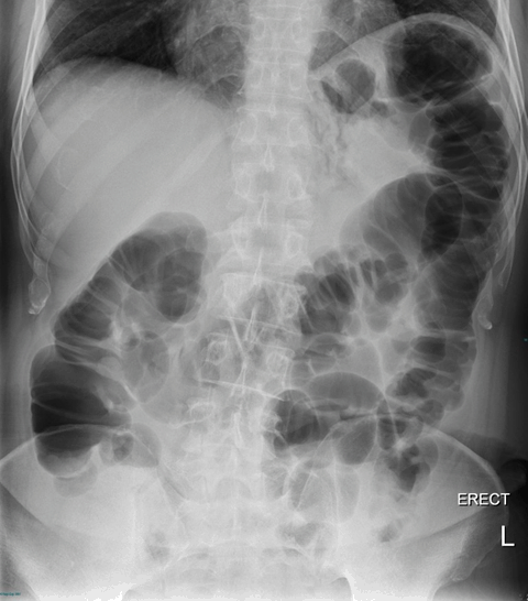
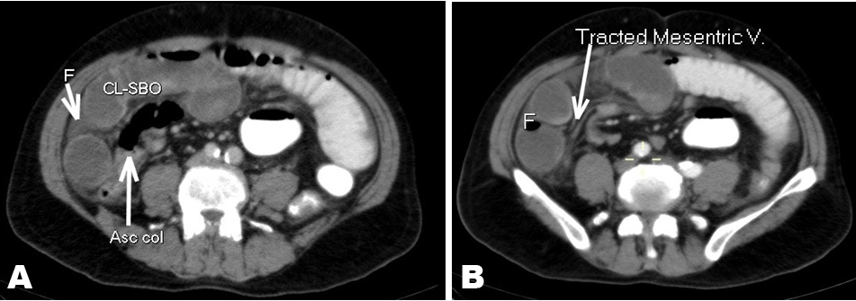
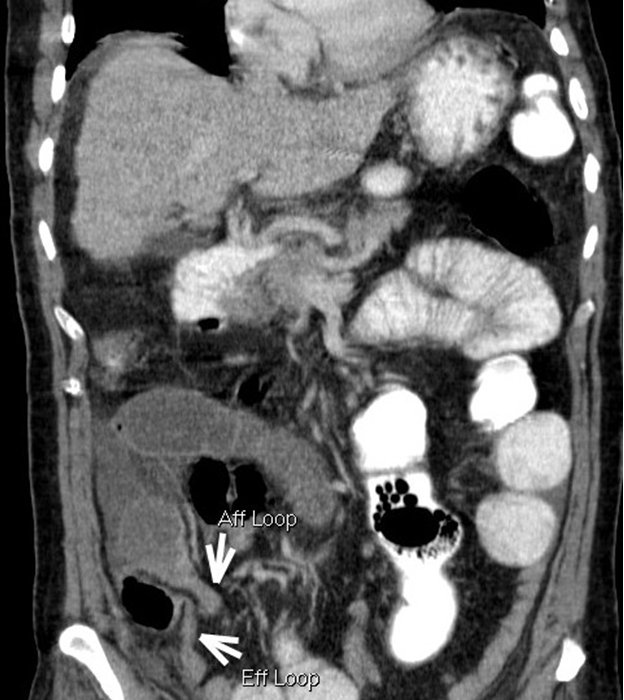
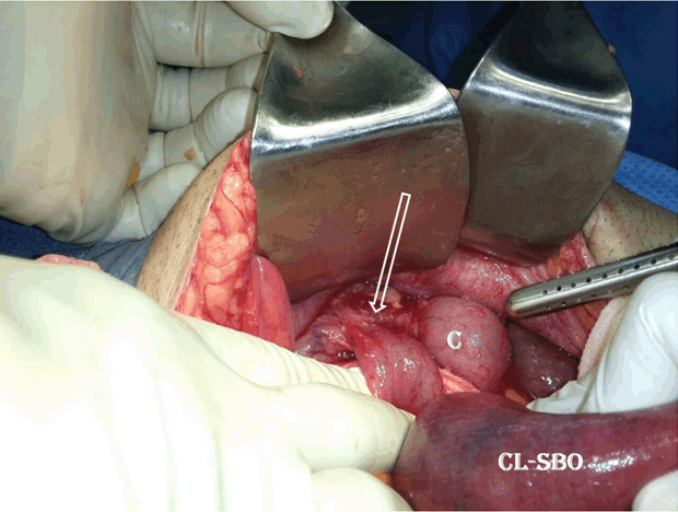
A 69-year-old male was presented to our emergency room with abdominal pain and distension the day after combined upper and lower gastrointestinal screening endoscopy done to investigate recurrent nausea, dyspepsia and episodic vomiting. There was no history of altered bowel habits. The patient denied any history of previous abdominal trauma. He had appendicectomy done thirty years ago. Physical examination revealed abdominal distension, right iliac fossa tenderness and hyperactive bowel sounds. The laboratory workup for the patient was unremarkable. A provisional diagnosis of intestinal obstruction was made. Plain radiography showed gaseous distension of the colon with no significant air-fluid levels (Figure 1). Contrast-enhanced multidetector computed tomography, with positive oral contrast preparation, was done for further assessment. Portal venous phase scans, revealed locally crowded fluid-filled dilated ileal segments within the right lower abdominal quadrant lateral to the antero-medially displaced cecum and right colon. There was a thin rim of reactive intra-peritoneal fluid collection detected in between these dilated loops (Figure 2A). Convergence of the related mesenteric vascular pedicle with marked stranding of surrounding mesenteric fat was noted, inferring prominent mesenteric congestion (Figure 2B). A peaking transitional zone was noticed just proximal and distal to these dilated fluid-filled segments. Moreover, the administered positive contrast in upstream bowel was absent in the dilated bowel segments and more distally. Coronal reformatted images nicely demonstrated the afferent and efferent limbs of this C-shaped dilated ileal loop lateral to the displaced cecum (Figure 3). There was no imaging sign of bowel perforation, necrosis, or ischemia. Based on these imaging findings; an extra-luminal bowel compression was postulated and a closed loop small bowel obstruction (CL-SBO) was presumptively diagnosed. The patient urgently underwent an exploratory laparotomy. A dusky dilated ileal loop; about 100 cm in length; was found residing in the right paracolic gutter and trespassing underneath an adhesive parieto-cecal peritoneal band, interconnecting the medially displaced cecum and right lower lateral abdominal wall (Figure 4). The band was released. Gradual normal pink color of the herniated ileal loops was restored and bowel salvage procedure was possible. The right colon and cecum were returned to their lateral anatomic position. The patient had an uneventful postoperative recovery. | ||||||
| ||||||
| ||||||
| ||||||
|
DISCUSSION
| ||||||
|
Internal abdominal herniation represents the protrusion of an abdominal organ via a peritoneal, mesenteric and/or omental defect into another abdominal compartment [4]. The hernial defect is either a congenital aperture e.g. Winslow’s foramen and omental defects, or potentially acquired post-surgical defects e.g., post-Roux-en-Y anastomosis or any laparotomy with adhesive bands formation. Location-wise, internal hernias have been classified as para-duodenal hernias, the foramen of Winslow hernias, trans-mesenteric hernias, pericecal hernias, inter-sigmoid hernias, and para-vesical hernias [4]. Although internal hernias are uncommon in clinical practice, they are reportedly associated with intestinal obstruction. Internal hernias are ominous as they are concealed and more liable to strangulation [1][2]. A CL-SBO is a type of intestinal obstruction in which a segment of bowel is occluded at two points by a mechanical hinder that impedes its spontaneous decompression. It has to be prospectively recognized to decrease morbidity and mortality rates due to associated vascular compromise [5]. Preoperative diagnosis of internal hernia is challenging due to its rarity and a myriad of clinical features varying from transient mal-digestive symptoms relieved spontaneously or gravitationally, to the classic symptoms of intestinal obstruction including abdominal pain, nausea and vomiting [2]. Pericecal hernias represent about 6–12% of all internal hernias [2][6]. In pericecal hernias, an ileal loop usually passes through a mesocecal defect to reside in one of the pericecal peritoneal recesses with variable extension in the right paracolic gutter. Embryologically; variations of midgut rotation, between 16–20 gestational weeks, result in formation of peritoneal recesses around the cecum [7]. The most common are the cecal fossa, cecal recess, retrocecal recess as well as the superior and inferior ileocecal recesses [7]. However, other potential acquired pericecal peritoneal recesses have been described, post-surgically as a consequence of adhesions [2][6]. Multidetector computed tomography role in evaluating small bowel obstruction is well established [8]. In view of its multiplanar reformat (MPR) capabilities it exhibits intra-luminal as well as extra-luminal causes of bowel obstruction, shows the mesenteric vasculature, peritoneum, abdominopelvic organs and depicts SBO complications [3][8]. Moreover; it is fast, increasingly available and becoming more accessible. Currently; it is the imaging modality of choice to document and assess internal hernias [9]. On MDCT, internal hernias are detected as cause of small bowel obstruction (SBO) by recognizing aggregated dilated bowel loops (hernia sac) along with pull up of associated mesentery and mesenteric vessels towards the hernia orifice as well as displaced adjacent structures within the abdominopelvic cavity [2][9]. In our case, MDCT revealed a combination of: (a) few fluid-filled dilated ileal segments in right lower abdominal quadrant, (b) local distortion of normal anatomic topography of bowel distribution with U-shaped or C-shaped arrangement of these loops on oblique coronal projections, (c) swirl of the related mesenteric fat and vessels, (d) reactive inter-loop fluid, and (e) abrupt change of bowel caliber (transitional zone) before and after these segments. The juxtaposition of contrast-filled proximal bowel, collapsed non-opacified distal bowel and fluid-filled dilated loops in between alerted us to the presence of CL-SBO. Moreover, the medial displacement of the cecum and right colon raised the suspicion of pericecal internal herniation as the most potential etiology as described in published literature reports [2][10]. Our case is distinguished by the location of the hernial orifice formed by a parieto-cecal acquired post-appendicectomy peritoneal adhesion interconnecting the anterior surface of the cecum medially, and right lower abdominal wall laterally. This finding has not described before, to the best of our knowledge, in the English literature. Association of internal hernias with gastrointestinal endoscopy has been previously reported [11]. Purgative-induced hyper-peristalsis in the preparation stage and gaseous distension during procedure have been postulated as forerunners [12]. Surgery is almost always the treatment of choice for small bowel obstruction caused by a pericecal hernia. Recently, laparoscopic adhesiolysis had been advocated and proved to be useful for both the diagnosis and treatment of small bowel obstructions [13]. | ||||||
|
CONCLUSION
| ||||||
|
In conclusion, post-appendicectomy adhesions may result in acquired potential peri-cecal recesses which can result in small bowel obstruction. Cecal displacement, mesenteric vascular distortion and imaging signs of closed-loop SBO on multidetector computed tomography should alert the interpreting radiologist to the possibility of pericecal hernia as an underlying cause. | ||||||
|
REFERENCES
| ||||||
| ||||||

|
[HTML Abstract]
[PDF Full Text]
|
|
Author Contributions
Mohamed Ragab Nouh – Substantial contributions to conception and design, Acquisition of data, Analysis and interpretation of data, Drafting the article, Revising it critically for important intellectual content, Final approval of the version to be published Ahmed Bassim Doma – Substantial contributions to conception and design, Acquisition of data, Analysis and interpretation of data, Drafting the article, Revising it critically for important intellectual content, Final approval of the version to be published Mohamed Ahmed Yousef Elshazly – Substantial contributions to conception and design, Acquisition of data, Analysis and interpretation of data, Drafting the article, Revising it critically for important intellectual content, Final approval of the version to be published Elsanousi Ibrahim Sabir – Substantial contributions to conception and design, Acquisition of data, Analysis and interpretation of data, Drafting the article, Revising it critically for important intellectual content, Final approval of the version to be published |
|
Guarantor of Submission
The corresponding author is the guarantor of submission. |
|
Source of Support
None |
|
Conflict of Interest
Authors declare no conflict of interest. |
|
Copyright
© 2018 Mohamed Ragab Nouh et al. This article is distributed under the terms of Creative Commons Attribution License which permits unrestricted use, distribution and reproduction in any medium provided the original author(s) and original publisher are properly credited. Please see the copyright policy on the journal website for more information. |
|
|