| Table of Contents | |
|
Original Article
| ||||||
| Usefulness of N-butyl cyanoacrylate embolization versus coil embolization for control of massive hemorrhage in patients with pelvic fracture | ||||||
| Zenjiro Sekikawa1, Izumi Torimoto1, Shigeo Takebayashi1, Hiroshi Manaka2, Kyota Nakamura3, Naoto Morimura3 | ||||||
|
1The Departments of Diagnostic Radiology, Yokohama City University Medical Center.
2The Neurosurgery, Yokohama City University Medical Center. 3The Critical Care and Emergency Center (KN, NM), Yokohama City University Medical Center. | ||||||
| ||||||
|
[HTML Abstract]
[PDF Full Text]
[Print This Article]
[Similar article in Pumed] [Similar article in Google Scholar] |
| How to cite this article |
| Sekikawa Z, Torimoto I, Takebayashi S, Manaka H, Nakamura K, Morimura N. Usefulness of N-butyl cyanoacrylate embolization versus coil embolization for control of massive hemorrhage in patients with pelvic fracture. Edorium J Radiol 2016;2:1–11. |
|
Abstract
|
|
Aims:
To evaluate the usefulness of N-butyl cyanoacrylate (NBCA) in transarterial embolization (TAE) in patients with pelvic fracture and massive hemorrhage.
Methods: We retrospectively reviewed 91 patients with pelvic fracture and massive hemorrhage requiring a >2 L of blood transfusion and TAE at a single institution from July 2008 to September 2015. The backgrounds and outcomes were compared between the NBCA group, which consisted of 46 patients treated by TEA including NBCA-Lipiodol embolization beginning in 2012, and the remaining 46 patients (non-NBCA group). Statistical analyses were performed to compare various factors among the two groups and to determine factors associated with mortality. Results: Between the NBCA group and the non-NBCA group, there were no significant differences in the backgrounds, the mean number of embolized arteries (4.22 ± 2.26 versus 4.24 ± 1.75), the mean time required for TAE (73.4 minutes ± 31.0 versus 74.3 minutes ± 29.2), or the mean cost of devices for TAE ($1535.7 ± 809.0 versus $1682.1 ± 914.3). The success rate of hemostasis by TAE and the mortality rate at 2 days or 30 days for the NBCA group and the non-NBCA group were 86.7%, 11.1%, 15.6% and 56.5%, 41.3%, 47.8%, respectively. Transarterial embolization without NBCA was a significant factor associated with 2-day and 30-day mortality. Conclusion: Transarterial embolization with NBCA could be used in resuscitative strategies for patients with pelvic fracture and massive hemorrhage because it is effective for improving early hemorrhage control and prognosis without increasing the cost of devices for TAE. | |
|
Keywords:
Cost-effectiveness, Cyanoacrylate, Embolization, Hemorrhage, Pelvis, Trauma
| |
|
Introduction
| ||||||
|
Pelvic fractures in trauma patients could be a major source of bleeding with subsequent hypovolemic shock. Hemodynamically stable patients had a mortality rate of 3.4%, and hemodynamically unstable patients had a mortality rate of 42% [1]. The management of unstable patients with pelvic fractures and massive hemorrhage is a controversial challenge requiring various specialties including a surgical team immediately available with a surgeon familiar with the principles of damage control and preperitoneal packing or an angiography suite with a skilled interventional radiologist for urgent transarterial embolization (TAE) of the internal iliac system with coils and gelatin absorbable sponges [2]. Transarterial embolization has many theoretical advantages and could be used by experienced individuals to treat multiple and anatomically distant bleeding sites from a single arterial access site [3]. Fangio et al.[1] reported that controlled resuscitation including vasopressor use and early pelvic TAE is effective for treating pelvic hemorrhage. N-butyl cyanoacrylate (NBCA), which almost immediately polymerizes upon contact with ionic fluids such as blood or with vascular endothelium, has been widely used as an embolic agent in the treatment of cerebrospinal arteriovenous malformations [4]. Recently, NBCA embolization was shown to be a safe alternative treatment for the management of gastrointestinal and postpartum hemorrhage [5][6]. The technique was found to stop arterial bleeding at various sites, even in cases when previous coil or particulate embolization failed. After we have had experiences with urgent NBCA embolization for the treatment of patients with those entities, we started to use the embolus to control massive hemorrhage in patients with pelvic fractures. The purpose of the present retrospective study was to describe our standardized techniques of NBCA-embolization and to assess the efficacy of embolization in the treatment of patients with pelvic fracture and massive hemorrhage with high mortality risks. | ||||||
|
Materials and Methods
| ||||||
|
Approval for this retrospective study was obtained from our institutional review board. We confirmed that the patients or the legal representatives of the patients in this study were given a comprehensive written statement of information about the clinical study, including information on TAE, and their consent was documented in the clinical records. One of the authors (I.T.) reviewed trauma database records and charts of TAE between September 2008 and September 2015 in the emergency department (ED) with a Level I trauma center of our hospital. There were 372 patients who underwent TAE for the control of hemorrhagic shock in the ED. Of those patients, 91 had pelvic fracture and massive hemorrhage requiring a > 2L blood transfusion. This study consisted of the 91 patients. The patients were between the ages of 18 and 84 years (48 males, 43 females) ; 47 patients had hemorrhagic shock from fall-related injuries, and 44 were victims of traffic accidents. The patients received fluid resuscitation consisting of the administration of an initial fluid bolus of lactated Ringer (2 L) over 15 to 20 minutes followed by ongoing lactated Ringers infusion (more than 250 mL/h) and packed red blood cells or fresh frozen plasma transfusions. After starting the fluid resuscitation, imaging examinations were performed as follows: an anteroposterior chest radiograph, a KUB radiograph and focused assessment with sonography for trauma examination (FAST) to detect hemoperitoneum, massive hemothorax, or hemopericardium. Patients in whom hypotension or tachycardia showed no response to the fluid resuscitation were transported directly to the angiographic suite in the ED with or without indwelling intra-aortic balloon occlusion catheter to maintain the blood pressure. In patients positive for FAST, surgical exploration and preperitoneal packing before TAE was selected to identify and control the abdominal bleeding sources because a surgical team and a TAE team were immediately available. Patients who transiently recovered from hypovolemic shock after the fluid resuscitation underwent multi-detector computed tomography (MDCT) scanning of the head and the trunk with a 16-row MDCT unit (Aquilion16; Toshiba Medical Systems, Ohtawara, Japan) before and after intravenous injection of contrast material. Beginning in 2015, all patients with pelvic fracture could undergo the scanning with an 80-row MDCT unit (Aquilion Premium; Toshiba Medical Systems, Ohtawara, Japan) which was equipped with a hybrid table for scanning and emergency treatment. Transarterial embolization was performed by one of two board-certified radiologists; S.T. or Z.S., who had embolization experience in the emergency department for 18 years and 8 years, respectively. Femoral arterial access using a 6-F introducer sheath (Radifocus II-H; Terumo, Tokyo, Japan) for angiography was usually performed at the contralateral site in more severely injured hemipelvises. First, a 5.2-F cobra occlusion catheter (Selecon MP catheter; Terumo) with a 0.035-inch guide wire (Radifocus; Terumo co.) was used to occlude the internal iliac arterial flow to achieve a rapid hemostasis. For selective catheterization of the internal iliac arterial branches, a 2.1-F microcatheter (Sniper 2, Terumo co.) was passed through the parent catheter into the branches of the arteries with a 0.016-inch microguidewire (Radifocus double angle, Terumo Co.) or a 0.014-inch microguidewire (Transend Floppy, Boston Scientific Co. Natick, Mass.). The ipsilateral internal iliac artery was catheterized with a 5-F shepherd Fhook catheter (Catex -CX, Gadelius medical co., Tokyo, Japan) or a 4-F cobra-head catheter (Terumo co.). A gelatin sponge (Spongel; Astellas Co., Tokyo, Japan) that was cut into 1–2 mm pieces and soaked in contrast medium (300 Img of iopamidol) was slowly infused under fluoroscopic control into the arteries responsible for bleeding to decrease the blood flow; the sponge was followed by a coil or NBCA embolization. Various coils used in TAE included 0.035 inch Tornado (Cook Co., Bloomington, Co.), 0.018 inch microcoils which were VorteX (Boston Scientific Co., Natick, MA) and C-stopper (Piolax Co., Yokohama, Japan). The microcoil with a diameter the same as the original diameter of the bleeding artery or 1–2 mm greater was positioned with the use of a pusher (C-stopper coil pusher, Piolax Co.). In addition to gelatin sponge particles and coils, NBCA (Histoacryl B, Braun, Melsungen, Germany) was used in use in the patients with pelvic fracture and massive hemorrhage beginning in 2012. NBCA was mixed with iodized oil (Lipiodol: Terumo Co.) at a ratio of 1:3 (25%) or 1:4 (20%) and was infused through a microcatheter with the tip located more than 3 cm distal to the parent catheter because the proximal overflow of the glue might cause occlusion of the artery at the level of the parent catheter tip. Following a flush of 20% glucose, a 2.5 mL-syringe containing 2.5 mL of a 20% mixture or 2.0 mL of 25% glue was connected to the microcatheter for the single-column injection technique. Under a maximum magnified fluoroscopic control, the operator continuously pushed the plunger with moderate pressure and kept pushing the plunger to avoid premature polymerization which caused a glue node in the microcatheter tip. The microcatheter was removed quickly when the operator observed the radiopaque material entered the target artery, the proximal back flow of the glue over the microcatheter tip or a glue node in the tip. The assistant for TAE immediately cleaned the withdrawn mirocatheter with a Lipiodol flush followed by saline flushes for once-repeated use using the inserter. When no back flow of blood was observed in the parent catheter after TAE, the parent catheter was removed and cleaned by a saline flush. The NBCA embolization of the internal iliac main trunk was performed with a different technique from that of other arteries with smaller calibers. The operator infused a 20% NBCA-Lipiodol mixture through a 2.4-F coaxial catheter of which the tip was placed in the anterior trunk. A second infusion of the glue was required when the glue cast was not observed in the proximal portion of the main trunk. The glue infusion into the internal iliac main trunk was performed through a parent catheter for a quick embolization when the patient had a risk of cardiac arrest with lower than 50 mmHg systolic pressure and bradycardia or ventricular fibrillation. Occlusion of the iliac artery and its branches was confirmed by non-selective angiography. However, the glue cast filling the target artery near the orifice was considered to be occluded without angiography. In patients in whom recurrent hemorrhagic shock occurred after the initial TAE achieved stabilized vital signs, a repeated TAE was performed. In patients in whom hemorrhagic shock was persistent after TAE, if possible, surgical repair was performed. The patients were divided into two groups. The NBCA group included patients in whom the glue embolization was performed in one or more arteries regardless of coil embolization in other arteries. The non-NBCA group included patients in whom all TAE was performed with only a coil or gelatin sponge. Patients' backgrounds were compared between two groups with regard to the mean values of age, injury severity score, serum level of hemoglobin and international normalized ratio (INR) as well as the systolic pressure and shock index ( heart rate/systolic pressure ) before TAE in the angiographic suit and the volume of blood transfusion before the end of TAE. The frequencies of patients with multi-compartmental hemorrhage, patients who underwent MDCT before TAE and patients with each type of Tile classification of pelvic fracture were compared [7]. We also compared the mean numbers of embolized arteries, the mean treatment time of TAE and the mean cost of the devices and embolic materials for TAE between the NBCA patients and the non-NBCA patients. The costs were calculated on the basis of costs equivalent to US$. The following costs were used: 21.2 $ per parent catheter, 112.5$ per parent catheter with occlusion balloon, 366.7$ per microcatheter, 20.0$ per guide wire for the parent catheter, 129.2$ per guidewire for microcatheter, 102.5$ per pushable coil, 147.5$ per pusher wire for the deployment of coil, 6.7$ per sheet of gelatin sponge, 20.5$ per 0.5 mL of NBCA, 41.2$ per 10 mL of Lipiodol. Clinical success in hemostasis was defined as cessation of angiographic extravasation resulting in the clinical cessation of bleeding and stabilization of vital signs. We documented the success rate by surgical repair after TAE and TAE intervention which included TAE alone, TAE after packing, TAE after both packing and surgical repair of injured bowels or solid organs. Each success rate, 2-day and 30-day mortality rate as well as the rate of comorbidities was compared between the NBCA group and the non-NBCA group. Statistical analyses were performed using the Excel add-on software, Xlstat (Addinsoft, Cologne, Germany). Quantitative variables were compared with the Mann-Whitney U test, and Pearson's chi square test was used to compare the categorical variables. We performed a multivariate analysis with a multiple logistic regression for binary dependent variables to determine the significant factors associated with the 2-day and 30-day mortality. In those analyses, a p-value less than 0.05 was considered statistically significant. | ||||||
|
Results | ||||||
|
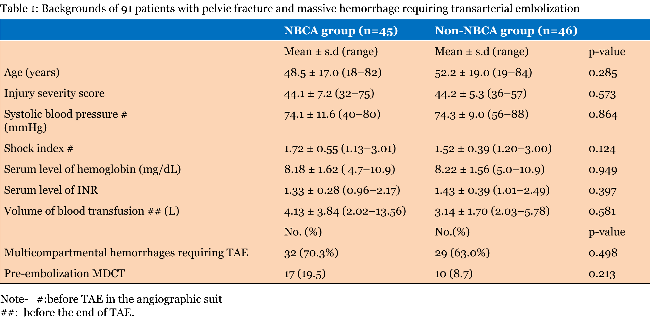
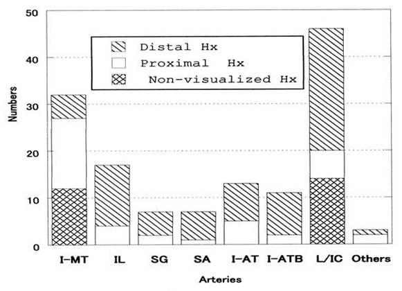
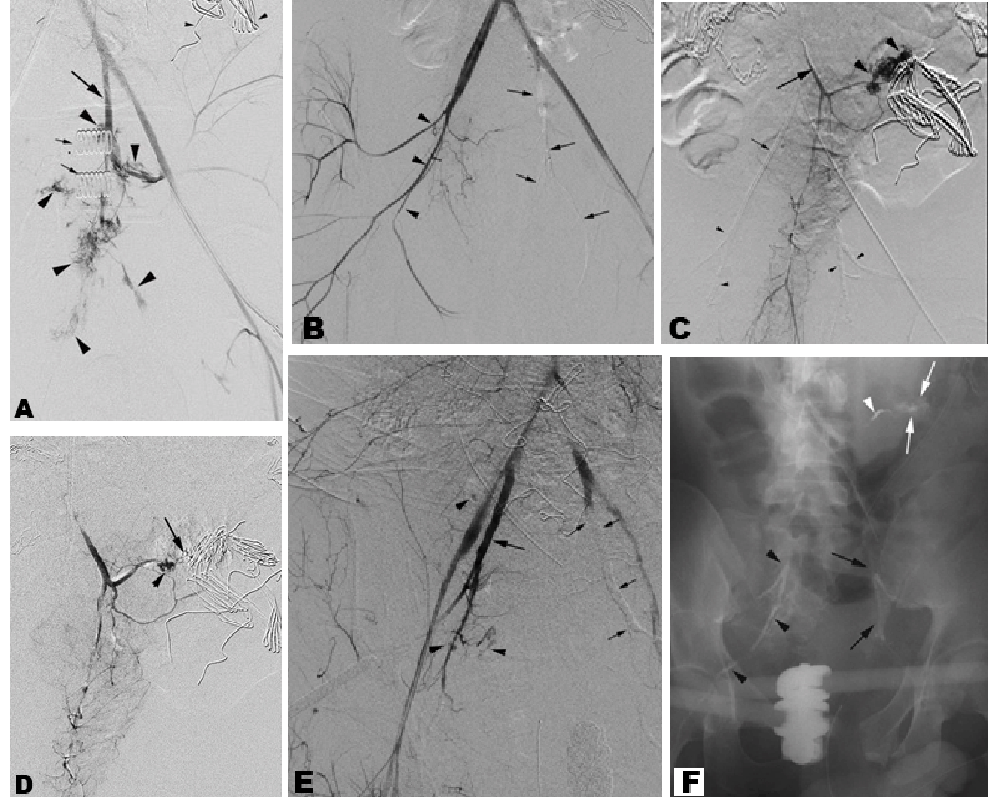
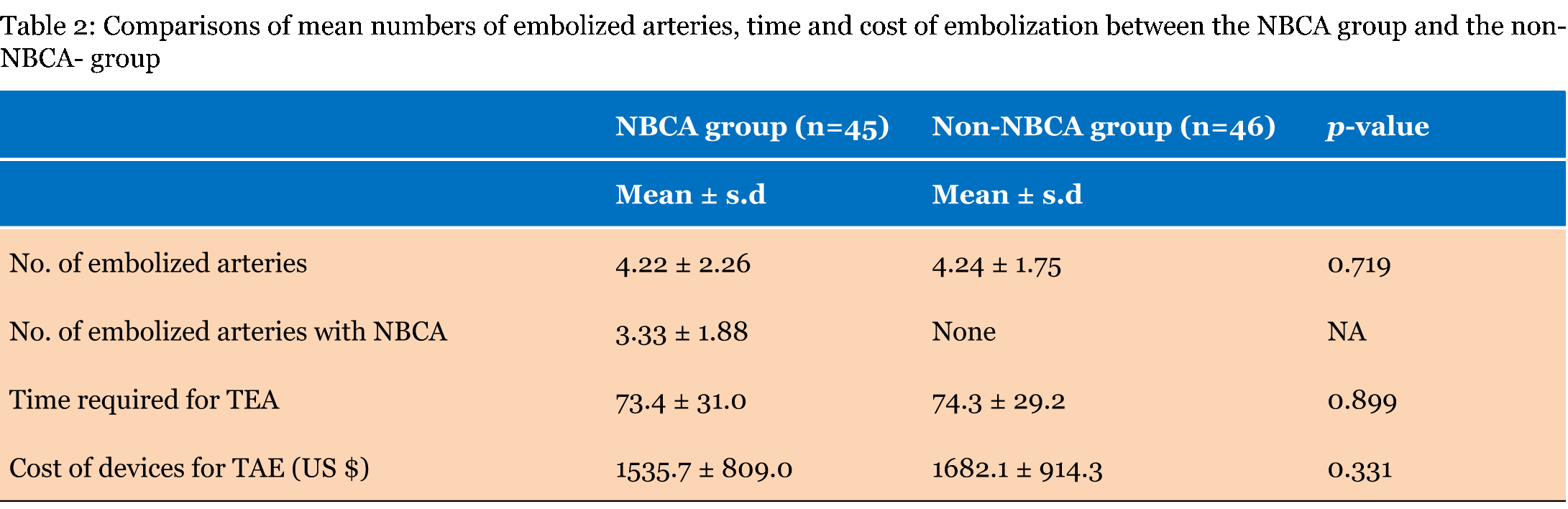
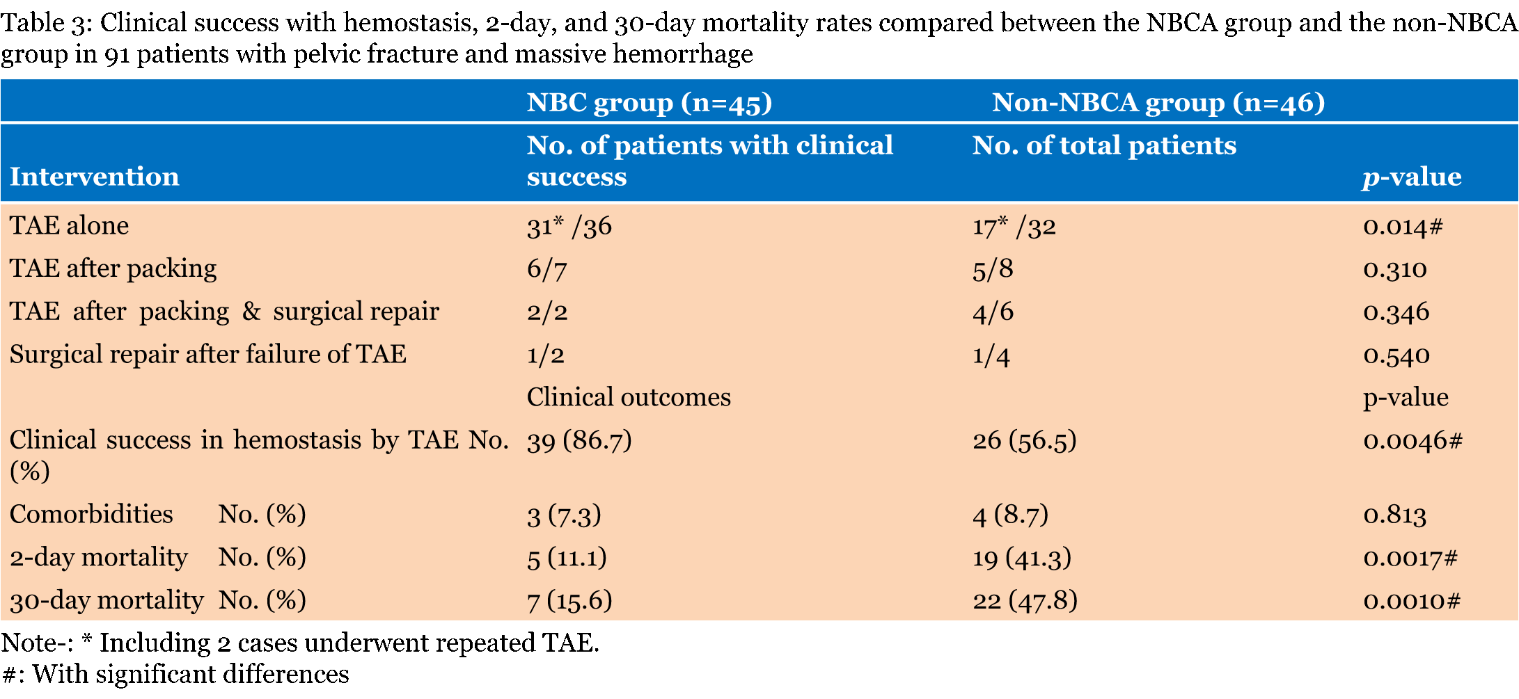
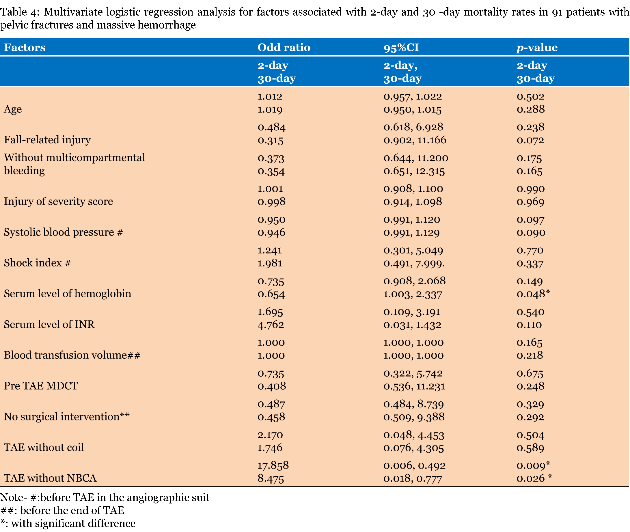
There were no significant differences in the mean values of the background data between the NBCA group and the non-NBCA group (Table 1). Tile type 1, type 2 and type 3 pelvic fracture patterns in the NBCA group were observed in 6 patients (12.2%), 31patients (70.7%), and eight patients (17.1%), respectively. Each fracture pattern in the non-NBCA group were seven patients (15.2%), 30 patients (65.2%) and nine patients (19.6%), respectively. There was no statistically significant differences in the frequencies of each type fracture between the two groups (p = 0.797, 0.710, and 0.827, respectively). Multi-compartmental hemorrhages requiring TAE in each NBCA group and non-NBCA group involved the retroperitoneum in 20 cases and 18 cases, the kidney in five cases and four cases, the adrenal gland in one case and two cases, the liver in four cases and three cases, the spleen in two cases and four cases, the intestine in two cases and one cases, the neck in three cases and two cases, and the extremity in three cases and two cases, respectively. Pre-embolization MDCT could be performed in 10 cases each of the NBCA group and the non-NBCA group because their hemodynamic stability was transiently recovered from hypovolemic shock. Seven cases in the NBCA group underwent the scanning equipped hybrid table for scanning and emergency treatment although their hemodynamically instabilities persisted. In the NBCA group, a total of 190 arteries were embolized. Of the 190 arteries, 136 (71.6%) were embolized with the NBCA-Lipiodol mixture. There were 73 (53.7%) arteries with distal hemorrhage and 37 (27.2%) with proximal hemorrhage. No visualized hemorrhage was shown in the remaining 26 arteries (19.1%) (12 main internal iliac trunk with spasm and 14 lumbar arteries with potential collaterals to the hemorrhagic arteries). The most frequent artery embolized with the NBCA-mixture was the lumbar or intercostal artery group (33.8%) followed by the internal iliac main trunk (23.5%), the iliolumbar artery (12.5%) and the internal iliac anterior trunk (9.6%) and the branch of the anterior trunk (8.0%) (Figure 1). Double sessions of 1.2–2.0 mL glue infusions through a 2.4-F microcatheter were required for the occlusion of the main internal iliac trunk without spasm (Figure 2A). Although a single infusion of the glue was able to make the glue cast filling 10 trunks with spasm, recanalization was observed in 8 (66.7%) of the 10 trunks; recanalization occurred in 6 trunks at the end of the TAE (Figure 2E) and in two trunks 3–4 hours after the first session of TAE. Each re-canalized internal iliac main trunk was completely occluded with a re-infusion of the glue. The remaining cases were two patients who underwent glue embolization through the parent catheter because the patients had a risk of cardiac arrest. The internal iliac artery could be sufficiently filled with the glue although no migration of the glue into the external iliac artery occurred; there was occlusion near the orifice of the artery. Compete occlusions of the lumbar or intercostal arteries were safely accomplished by an infusion 0.2–0.3 mL of a NBCA-Lipiodol mixture. The glue embolization was not performed in two hemorrhagic lumbar arteries that were connected to the Adamkiewicz arteries, but in both upper and lower lumbar arteries with a collateral connection to the peripheral arterial branch of the responsible artery. The remaining 12 embolized lumbar arteries without visualized hemorrhage were the fourth lumbar arteries with collaterals to injured iliolumbar arteries. The sacral arteries embolized with the glue included one medial sacral artery. Four glue embolized arteries that were assigned to others were two the inferior phrenic arteries, one inferior adrenal artery and one colonic arterial branch which was embolizd with a combination of coil (Figure 3C-D). Other arteries treated with a combination of glue and coil were two internal iliac anterior trunks in which the glue was used in the treatment of extravasation caused by deployed coils. The non-NBCA group had 231 embolized arteries which were 191 occluded by gelatin-sponge particles followed by coil deployments and 40 arteries embolized with gelatin-sponge particles alone. There were no statistically significant differences in the mean number of embolized arteries, the mean time required for TAE, or the mean cost of TAE between the NBCA group and the non-NBCA group (Table 2). The clinical success rate in hemostasis by TAE only was 39 patients (86.7%) in the NBCA group and 26 patients (56.5%) in the non-NBCA group, with a statistically significance difference between the groups (p = 0.0046) (Table 3). The patients with success in each group included 2 patients in whom repeat TAE was performed. Surgical repairs after TAE failure achieved hemostasis in one of two patients in the NBCA group and in one of four patients in the non-NBCA group. Major complications were limited to gluteal muscle necrosis in one patient in each group. The patients underwent bilateral internal iliac artery with glue in the NBCA group and gelatin sponge and coils in the non-NBCA group. The patients survived although their hospitalizations were prolonged. TAE of the internal iliac main trunk caused no migration of the glue in the external iliac artery. Transarterial embolization of the lumbar or the intercostal artery also did not cause glue migration in the aorta because the parent catheter was wedged into a lumbar artery. There was no complication of entrapment of the catheter within the embolized artery although the parent catheter always occluded by refluxed glue from the microcatheter in TAE of the lumbar and the intercostal artery or TAE of the other arteries in which the distance between the parent catheter and the microcatheter was not far enough to prevent it from reaching glue backflow. In the present TAE series with pelvic fracture with massive hemorrhage, the 2-day mortality and 30-day mortality rates for the NBCA group and the non-NBCA group were 11.1%, 15.6% and 41.3%, 47.8%, respectively (Table 3). The multivariate analysis showed that TAE without NBCA was a significant factor associated with the 2-day mortality and 30-day mortality (odds ratio; 17.868, 8.475 respectively and p = 0.009, 0.026, respectively). (Table 4). The serum level of hemoglobin was also a significant factor in associated with the 30-day mortality (odds ratio; 0.654 and p = 0.048). | ||||||
| ||||||
| ||||||
|
| ||||||
|
| ||||||
|
| ||||||
|
| ||||||
| ||||||
| ||||||
| ||||||
|
Discussion
| ||||||
|
Patients with pelvic fracture and massive hemorrhage are thought to have a high mortality risk when their hypotension does not respond to a transfusion [8]. The appropriate management of massive hemorrhage due to pelvic fracture and retroperitoneal or abdominal trauma remains a large challenge for emergency medicine because more than half of patients with pelvic fracture had also other compartmental hemorrhage as shown in this study. The management of patients with polytrauma requires a multidisciplinary team because prompt restoration of hemodynamic stability is important. Pre-peritoneal packing could be used if the surgical team has the appropriate knowledge and experience. However, surgical intervention seems to be less effective in the management of fracture-related retroperitoneal hemorrhage [9]. Instead, TAE with gelatin-sponge particles and coils is reported to be an effective method for controlling retroperitoneal hemorrhage caused by fracture-associated retroperitoneal arterial hemorrhage [8]. The mechanism of hemostasis by gelatin sponges or coils involves thrombus formation, which requires the patient's hemostatic capacity for successful embolization. However, severe coagulopathy, which is frequently associated with life-threatening massive hemorrhage in patients with pelvic fracture, might prohibit thrombus formation and, thus, the achievement of hemostasis might fail. NBCA is permanent embolic material and works independently of the patient's hemostatic capacity and is experimentally superior to gelatin-sponge and coils [10]. Kish et al. [11] reported that NBCA was successful in controlling active extravasation of contrast medium in 10 patients in whom standard coil and particulate embolization had failed. The advantages of TAE with NBCA over coil and gelatin sponge particles include its high success rate for occlusion by single infusion in most arteries other than internal iliac main trunk, its effectiveness for controlling bleeding in severe coagulopathic conditions and the high durability of embolization [6] [11]. Glue embolization is also useful in pseudoaneurysm associated with trauma because the lesion could be completely filled with the infused glue [12]. Glue embolization with these benefits provides a prompt restoration of hemodynamic stability in patients with pelvic fracture, multi-compartmental injuries and massive hemorrhage. NBCA embolization is also superior to gelatin-sponge particles and coil embolization in terms of costs if the catheter infused with the NBCA-Lipiodol mixture is cleaned and repeatedly use. Conversely, the method is inferior to gelatin-sponge and coil embolization in cost if the infused catheter is limited to a single use. Cleaning of the catheter is more effective by Lipiodol followed by infusion of gelatin-sponge particles than Lipiodol alone [13]. A larger caliber-microcatheter is more easily cleaned. However, glue embolization with mirocatheters did not shorten the treatment time compared with TAE with gelatin sponge particles and coils because the time is mainly dependent on the time for the placement of the microcatheter into the injured arteries. TAE with NBCA infused through the parent catheter was reported to shorten the treatment time [6]. However, glue embolization with mirocatheters did not shorten the treatment time compared with TAE with gelatin sponge particles and coils because the time is mainly dependent on the time needed for the placement of the microcatheter into the injured arteries. Embolizaton with NBCA has been thought to be more technically challenging than embolization with microcoils, and requires specific training and expertise [14]. A major reason for this challenge is that it is difficult to prevent premature polymerization of NBCA that might cause insufficient embolization and proximal overflow and a potential risk of the entrapment of catheter within the embolized artery. NBCA is usually mixed with Lipiodol at ratios of 1:1 to 1 :10 depending on the distance from the microcatheter tip to the bleeding site and on the velocity of blood flow [6]. A simplified technique maintaining some of the steps, as used in this study, would result a safe and successful NBCA embolization. There is no risk of the entrapment of a microcatheter by a 20 % or 25% NBCA-Lipiodol mixture which is infused through a microcatheter. The procedure should be finished within several seconds after the operator observes a flow of the glue from the microcatheter tip under a maximum magnified fluoroscopic control because Lipiodol makes the mixture radiopaque and increased 25% mixture polymerization time is only 4 seconds [15]. Continuously pushing the plunger filled with high viscosity glue is one of the most important steps to prevent premature polymerization in the tip of the microcatheter. A quick removal of the microcatheter is also important after the injection is stopped when the operator observes the node and opacification of the target artery with the glue and the proximal back flow of the glue over the microcatheter tip. The position of a parent catheter is also important because the artery at the level of its tip is frequently occluded by the proximally refluxed glue or a large glue node in the microcatheter tip. In contrast to small or medium sized arteries, it is difficult to embolize the internal iliac main trunk with the NBCA mixture. First, a double session of the infusion of the mixture is required through a non-bolus infusion into a 2.1-F or 2.4-F mirocatheter. Premature polymerization with a large node of the glue should be avoided especially in the embolization of the trunk because the node would migrate in the external iliac artery or the femoral artery upon the removal of the parent catheter. A prompt and complete occlusion of the trunk is required for a bolus infusion through a parent catheter. However, this technique is limited in use because of the fear of cardiac arrest because it has a high risk for migration of the glue into the external iliac system. Second, the operator should pay attention to the injection speed in the main trunk with spasm in patients with an indwelling IABO catheter with or without inflated balloon because the infused glue is easily refluxed in the trunk with a slow flow velocity. In the present series of pelvic fracture with massive hemorrhage, NBCA embolization caused no serious complications other than gluteal muscle necrosis in one case in whom the bilateral internal iliac main trunk with spasm was occluded. An infusion of a 20% NBCA mixture could reach the arterial branches 300 micrometers in diameter or smaller, but caused no ischemic change to the arteries [16]. The necrosis in patients with pelvic fracture is thought to be partially a result of blunt trauma to the muscles and mostly the result of the ischemia from TAE [17] because most of cases who underwent embolization bilateral internal iliac main trunks with either a NBCA mixture, gelatin-sponge or coils experienced no gluteal muscle necrosis with no predominant occurrence of necrosis among the embolic materials. This present study does have limitations. Whether glue embolization was used in patients with pelvic fracture and massive hemorrhage was not based on randomization. The survival rate in our series made it difficult to compare patients who had severe comorbidities with those who did not. Resuscitation technique other than TAE might have improved after 2012 when NBCA embolization was introduced in our institution. Furthermore, eight recent cases that underwent NBCA embolization also underwent pre-embolization MDCT with hybrid table for scanning and emergency treatment. Despite the limitations of the present study, TAE with NBCA could be used in resuscitative strategies for patients with pelvic fracture and massive hemorrhage because the technique is effective for improving early hemorrhage control and prognosis without increasing the cost of devices for TAE. | ||||||
|
References
| ||||||
| ||||||
|
[HTML Abstract]
[PDF Full Text]
|
|
Author Contributions
Zenjiro Sekikawa – Substantial contributions to conception and design, Acquisition of data, Analysis and interpretation of data, Drafting the article, Revising it critically for important intellectual content, Final approval of the version to be published Izumi Torimoto – Analysis and interpretation of data, Revising it critically for important intellectual content, Final approval of the version to be published Shigeo Takebayashi – Analysis and interpretation of data, Revising it critically for important intellectual content, Final approval of the version to be published Hiroshi Manaka – Analysis and interpretation of data, Revising it critically for important intellectual content, Final approval of the version to be published Kyota Nakamura – Analysis and interpretation of data, Revising it critically for important intellectual content, Final approval of the version to be published Naoto Morimura – Analysis and interpretation of data, Revising it critically for important intellectual content, Final approval of the version to be published |
|
Guarantor of submission
The corresponding author is the guarantor of submission. |
|
Source of support
None |
|
Conflict of interest
Authors declare no conflict of interest. |
|
Copyright
© 2016 Zenjiro Sekikawa et al. This article is distributed under the terms of Creative Commons Attribution License which permits unrestricted use, distribution and reproduction in any medium provided the original author(s) and original publisher are properly credited. Please see the copyright policy on the journal website for more information. |
|
|